Advertisement

For more than a year we have been conducting intensive research and development in the field of producing training models using 3D printers.
Newly developed materials and printing processes make it possible to design the bony and soft tissue parts so realistically that animal specimens can now be completely dispensed with as a training base.
For you as the organiser of various surgical courses, this means no time consuming procurement and disposal of any animal cadavers and a pleasant training atmosphere for the participants.
Further advantages result from the human anatomy of the teeth and jaws and the possibility of fitting these models into the most common phantom heads.
The bony parts of each model are always divided into cortical and cancellous bone, so they always have a very natural feel and a very good insertion torque when drilling or in piezo surgery.
Furthermore, we are able to produce the bone in different qualities between D1-D4.
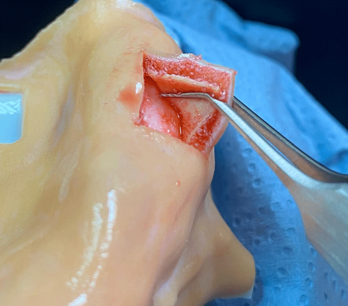
The biggest challenge was to design the soft tissue in such a way that it is fixed to the bone but can still be lifted off very naturally with instruments.
Flap techniques can also be taught impressively on these models by slitting the periosteum, and the removal of grafts from the palate can be carried out very realistically with recession coverage using the tunnelling technique.
All our models can be customised with your company logo.
The table mounting system (wet-lab-station) developed by us brings all our models into a natural patient position in all your courses.
The integrated mobile phone holder makes it possible for course participants to film their training on the model so that the course can be rewatched.
The speaker-station is also equipped with a high-quality 4K camera for transmission to a projector or television.

Atterseestraße 5
4863 Seewalchen am Attersee
Austria
4863 Seewalchen am Attersee
Austria
more information
Hall 11.3 | A080

